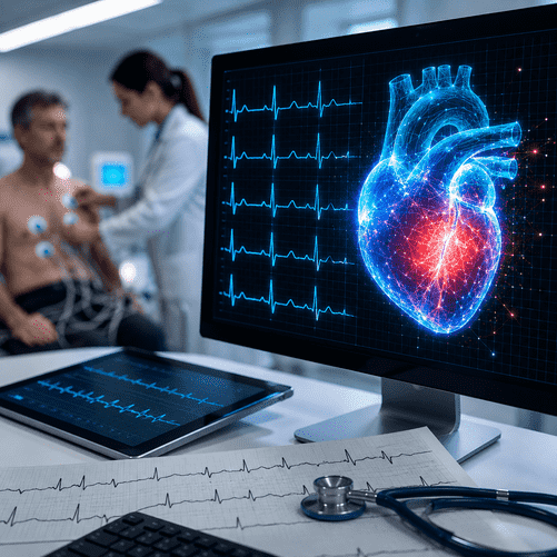
A system trained on more than 10 million electrocardiograms can uncover signs of heart failure and valve disease invisible to the human eye, potentially transforming one of medicine’s most common tests into a powerful early-warning tool.

One of the oldest diagnostic technologies in modern medicine is being given an extraordinary new capability.
Researchers from Imperial College London have developed an artificial-intelligence system capable of analysing a routine electrocardiogram and detecting hidden signs of serious heart disease in less than two seconds.
The technology was presented at the European Society of Cardiology Congress in Munich and could eventually allow hospitals to identify patients at high risk of heart failure or heart-valve disease almost immediately after an ECG is recorded.
An ECG normally takes only seconds to perform. Electrodes placed on the chest record the electrical signals produced as the heart beats, generating the familiar wave-like pattern doctors use to diagnose abnormal rhythms, heart attacks and other cardiovascular problems.
But the new research suggests those electrical traces contain considerably more information than physicians can recognise visually.
Artificial intelligence can apparently see it.
The system has been trained to identify subtle combinations of electrical activity associated with structural abnormalities inside the heart — including weakening of the heart’s main pumping chamber and disease affecting its valves.
If the technology performs equally well in real-world clinical use, it could change how millions of patients are screened.
Finding Disease Hidden in Plain Sight
The breakthrough stems from an increasingly important area of medical AI: extracting diagnostic information from tests that hospitals already perform routinely.
Researchers trained the system using 10.6 million ECGs, together with clinical reports describing the patients’ conditions.
A more targeted training dataset included 72,475 ECGs linked to echocardiogram results, allowing the algorithm to learn relationships between electrical patterns and physical changes in the heart.
The resulting AI was then tested on two separate groups of US hospital patients.
In the larger group of more than 61,000 patients, the algorithm correctly identified about 81% of people with reduced heart-pumping function, an important form of heart failure.
It also detected patients with aortic stenosis — a narrowing of one of the heart’s principal valves — with performance that varied between study groups. In the smaller validation cohort, it correctly identified approximately 90% of patients with the condition.
Those numbers do not mean the AI can replace a cardiologist or an echocardiogram.
Researchers emphasise that it cannot provide a definitive diagnosis by itself.
Its potential value lies elsewhere: triage.
A patient whose ECG appears normal to a physician but is flagged as high-risk by the AI could be moved rapidly to further investigation.
That next step would usually involve an echocardiogram, an ultrasound scan that shows the heart’s chambers, valves and pumping function in detail.
Why Speed Matters
Heart disease remains one of the world’s leading causes of death, and delays in diagnosis can have serious consequences.
Heart failure often develops gradually. Symptoms such as fatigue, breathlessness and swollen ankles can initially be attributed to ageing, respiratory illness or reduced fitness.
Heart-valve disease can be similarly difficult to recognise before it becomes advanced.
The problem is not always the absence of a diagnostic test.
It is access.
Echocardiography requires specialist equipment and trained staff, and patients in overstretched health systems can sometimes wait weeks or months for scans.
The AI-powered ECG could help doctors decide who needs those scans most urgently.
Professor Fu Siong Ng of Imperial College London, who oversaw the research, said the technology could identify people at greatest risk and allow hospitals to prioritise them for echocardiography.
That could have a powerful practical effect.
Instead of sending every patient with uncertain symptoms through the same diagnostic pathway, hospitals could potentially use a cheap and widely available ECG to identify the highest-risk cases first.
One Billion ECGs a Year
The potential reach of the technology is unusually large because the ECG is already embedded throughout global healthcare.
Approximately one billion ECGs are performed worldwide each year, according to the British Heart Foundation.
They are used in emergency rooms, hospital wards, outpatient clinics, ambulances and family doctors’ surgeries.
Unlike many emerging diagnostic technologies, widespread adoption would therefore not require hospitals to build an entirely new testing infrastructure.
The intelligence is added largely in software.
That distinction is important.
Much of the excitement surrounding medical AI involves highly specialised imaging equipment or expensive genomic testing. An AI-enabled ECG could instead enhance a technology that has existed for more than a century.
The machine does not change.
The interpretation does.
A ‘Superhuman’ Layer of Analysis
Researchers describe the technology as capable of identifying information that is effectively hidden from conventional human interpretation.
That does not mean doctors are being replaced.
It means the algorithm is being used to recognise statistical patterns across millions of ECGs that no individual clinician could reasonably learn.
Dr Ahmed El-Medany, a British Heart Foundation clinical research fellow who led the analysis at Imperial, said the findings suggest that “far more information” may exist inside routine ECGs than doctors can detect simply by examining them visually.
This is a recurring pattern in medical AI.
Algorithms trained on sufficiently large datasets can sometimes recognise correlations too subtle or complex for conventional clinical interpretation.
Similar techniques are being investigated in radiology, pathology, dermatology and ophthalmology.
An eye scan designed to examine the retina, for example, may also contain clues about cardiovascular risk.
A chest X-ray may reveal patterns associated with diseases beyond the condition for which the scan was originally ordered.
The ECG is now joining that category.
The NHS Trial
The next stage will determine whether the impressive research results translate into routine healthcare.
The AI system is currently being evaluated using ECGs from 590 NHS patients in London and Bristol.
Researchers want to see whether the algorithm performs consistently in British patients and whether it can be integrated into real clinical workflows without generating excessive false alarms.
This is a critical step.
AI systems frequently perform extremely well during controlled retrospective studies but encounter problems when introduced into hospitals.
Different patient populations, different ECG machines, incomplete medical records and variations in clinical practice can all affect accuracy.
An algorithm that identifies too many false positives might increase rather than reduce pressure on cardiology departments.
Patients incorrectly flagged as high-risk could undergo unnecessary testing.
Conversely, missed cases would create a false sense of reassurance.
The real-world trials will therefore evaluate not merely whether the AI works mathematically, but whether it actually improves patient care.
Researchers estimate the technology could be approximately two years away from routine clinical use, assuming further studies are successful.
From Hospital Machines to Handheld Devices
Perhaps the most intriguing possibility lies beyond conventional hospitals.
Researchers are investigating whether the same diagnostic information can be captured using smaller, portable ECG devices.
Modern handheld ECG systems can already record heart rhythms using compact sensors, and some consumer smartwatches include basic electrocardiographic capability.
If highly sophisticated diagnostic AI could eventually operate on signals captured by cheaper portable equipment, cardiovascular screening could become dramatically more accessible.
A family doctor might perform a rapid ECG during an ordinary consultation.
An ambulance crew could identify signs of hidden heart failure before arriving at hospital.
Patients in rural or underserved regions could potentially receive sophisticated cardiovascular risk screening without immediate access to a specialist cardiology centre.
In the longer term, the combination of portable sensors and AI could move aspects of cardiac diagnosis much closer to the patient.
The Wider AI Medicine Revolution
The research presented in Munich reflects a broader transformation currently sweeping medicine.
Artificial intelligence is moving from administrative applications into direct clinical decision-making.
Algorithms are already being tested for detecting cancer on medical scans, analysing pathology slides, predicting deterioration in intensive-care patients and identifying abnormal heart rhythms.
What differentiates the Imperial research is the simplicity of the input.
A standard ECG is inexpensive, fast and extremely common.
Transforming that test into a screening tool for diseases that traditionally require ultrasound imaging could make AI useful at enormous scale.
The Imperial research team has also created a spinout company, Cardiovolt.ai, intended to help move the technology from the laboratory into hospitals.
The initial focus is expected to remain on identifying hidden heart failure and valve disease.
But the ambition is broader.
British Heart Foundation reporting says related AI models trained on ECG data are being investigated for signals associated with more than a dozen conditions, including high blood pressure, diabetes and kidney disease.
That raises an extraordinary possibility.
In the future, the ECG may no longer be regarded primarily as a test of the electrical activity of the heart.
It could become a general biometric window into human health.
Doctors Still Remain Central
Despite the enthusiasm, cardiologists are stressing that the technology is not intended to replace physicians.
The AI identifies risk.
Doctors determine what that risk means.
A suspicious ECG still needs to be interpreted in the context of symptoms, medical history, blood tests, imaging and physical examination.
The British Heart Foundation cautions that the system will not identify every patient with disease and cannot definitively diagnose or exclude heart failure or valve disorders on its own.
That limitation is crucial.
The most realistic future for medical AI is increasingly emerging as one of collaboration rather than substitution.
Machines can analyse enormous datasets and recognise subtle patterns.
Doctors provide clinical judgment, context and responsibility.
Used correctly, the combination may be considerably more powerful than either alone.
Reinventing a 120-Year-Old Test
The electrocardiogram has changed surprisingly little since the Dutch physiologist Willem Einthoven helped establish the modern ECG at the beginning of the 20th century.
The basic principle remains the same: measure tiny electrical changes produced by the beating heart and display them as a waveform.
Yet artificial intelligence may now be turning that familiar waveform into something fundamentally different.
Instead of telling physicians only how the heart is beating, the ECG could begin revealing information about how strongly it is pumping, whether its valves are narrowing and potentially whether disease is developing elsewhere in the body.
And the analysis can be completed almost instantly.
The innovation is therefore not simply another example of artificial intelligence performing a medical task faster.
It illustrates a potentially more significant transformation: AI allowing medicine to extract entirely new information from tests that doctors thought they already understood.
A diagnostic instrument invented more than a century ago may be about to acquire capabilities its creators could never have imagined.
And if ongoing NHS trials confirm the results, one of medicine’s simplest tests could become one of its most powerful early-warning systems.